Study of CSF flow physics and its parameters at the level of aqueduct in normal individuals
Abstract
Phase-contrast MRI (PC-MRI) is a rapid, simple, and non- invasive technique, and is sensitive to CSF flow. It has been available for some time and been used in the past decade in the evaluation of cranial and spinal CSF flow, demonstrating a mechanical ‘coupling between cerebral blood and CSF flows throughout the cardiac cycle and the temporal coordinated succession of these flows’ in normal people. The technique led to a better understanding of the pathophysiological basis of diseases with dysfunction of CSF flow.
The aim of the study is to study the physics of the CSF flow and to establish the normal parameters of the CSF flow at the level of the aqueduct. MRI brain with CSF flow study was done in 40 patients. These patients were in the age group of 20-60 years and came with no significant clinical complaints.
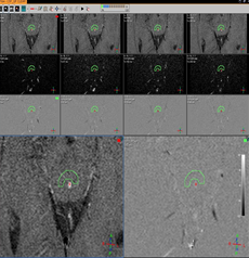
Phase-contrast MRI scanning was used following the CSF Quantitative flow protocol and CSF_ DRIVE protocol was followed. Forward flow volume, Backward flow volume, regurgitate fraction, absolute stroke volume, Stroke volume was calculated at the level of cerebral aqueduct provides the best understanding of CSF flow physics and normal CSF parameters.
The stroke volume of 55 % of individuals was seen in the range of 2.0 to 3.0 and 45% of individuals was seen in the range of 1.0 to 2.0 and Absolute stroke volume of maximum individuals i.e. 72.5% was seen in the range of the 3.6 to 4.5 ml/min.
Downloads
References
Guyton, Arthur C.; Hall, John Edward (2005). Textbook of medical physiology (11th ed.). Philadelphia: W.B. Saunders. p. 764. ISBN 978-0-7216- 0240-0.
Brian O, Tom P, Wang D, et al. relevance to cerebrospinal fluid physiology and therapeutic potential in hydrocephalus. Cerebrospinal Fluid Res 2010; 7:15.
Bradley WG. Magnetic resonance imaging in the evaluation of cerebrospinal fluid flow abnormalities. Man, Res Q 1992; 8:169-196
Wagshul, M.E., Eide, P.K. & Madsen, J.R. The pulsating brain: A review of experimental and clinical studies of intracranial pulsatility. Fluids Barriers CNS 8, 5 (2011).
Stoquart-El S, Sankari P, Lehmann C, et al. Phase-contrast MRI support for the diagnosis of aqueductal stenosis. J Neuroradiol 2009; 30:209- 214.
Stadlbauer A, Salamonie E, Brenneis C, et al. Magnetic resonance velocity mapping of 3D cerebrospinal fluid flow dynamics in hydrocephalus: preliminary results. Eur Radiol 2012;22(1):232-242. DOI10.1007/00330-011- 2247-7.
Yildiz H, Yazici Z, Hakyemez B, Erdogan C, Parlak M. Evaluation of CSF flow patterns of posterior fossa cystic malformations using CSF flow MRI. Neuroradiology 2006; 48:595–605
Bradley WG, Whittemore AR, Kirtman KE, Watanabe AS, Homyak M, Teresi LM, et al. Marked cerebrospinal fluid void: indicator of successful shunt in patients with suspected normal-pressure hydrocephalus. Radiology 1991; 178:459–66
Ng SE, Low AM, Tang KK, Lim WE, Kwok RK. Idiopathic normal pressure hydrocephalus: correlating magnetic resonance imaging biomarkers with clinical response. Ann Acad Med Singapore 2009; 38:803–8
Ball MJ, Dayan AD. Pathogenesis of syringomyelia. Lancet 1972; 2:799–801
Burgers P, Idy-Peretti I, Iffenecker C, Parker F, Jolivet O, Hurth M, et al. CSF flow measurement in syringomyelia. AJNR Am J Neuroradiol 2000; 21:1785–92
Shaw CM, Alvord EC Jr. Cava septic pellucid et vergae: their normal and pathological states. Brain 1969; 92:213–23.
McGirt MJ, Nimjee SM, Fuchs HE, George TM. Relationship of cine phase-contrast magnetic resonance imaging with outcome after decompression for Chiari I malformations. Neurosurgery 2006; 59:140–6
Buxton N, Macarthur D, Mallucci C, Punt J, Vloeberghs M. Neuroendoscopic third ventriculostomy in patients less than 1 year old. Pediatr Neurosurg 1998; 29:73–6
Henry-Feugeas MC, Idy-Peretti I, Blanchet B, Hassine D, Zannoli G, Schouman-Claeys E. Temporal and spatial assessment of normal cerebrospinal fluid dynamics with MR imaging. Magn Reson Imaging. 1993; 11:1107–1118.
Kolbitsch C, Schocke M, Lorenz IH, Kremser C, Zschiegner F, Pfeiffer KP, et al. Phase-contrast MRI measurement of systolic cerebrospinal fluid peak velocity (CSFV (peak)) in the aqueduct of Sylvius: a noninvasive tool for measurement of cerebral capacity. Anaesthesiology. 1999; 90:1546–1550.
Jeong Hyun Lee, MD, Ho Kyu Lee, MD, Jae Kyun Kim, MD Hyun Jeong Kim, MD, Ji Kang Park, MD, and Choong Gon Choi, MD CSF Flow Quantification of the Cerebral Aqueduct in Normal Volunteers Using Phase-Contrast Cine MR Imaging. Korean J Radiol. 2004 Apr-Jun; 5(2): 81–86.