Fibroepithelial hyperplasia of Gingiva: A report of two cases
Abstract
Introduction- Fibromas are the most common fibrous overgrowths of the oral cavity that occur in response to a low-grade stimulus. The treatment involves; efficient plaque control, removal of etiological factors, and conservative excision of the tissue to avoid recurrence. Hence, it is necessary that clinicians have adequate knowledge about such fibrous overgrowths so as to differentiate them from other similar appearing lesions.
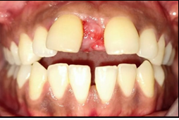
Case discussion- Two patients reported a chief complaint of overgrowth in the gums. On clinical examination, the overgrowths were diagnosed as Fibromas. Radiographic and hematological investigations were carried out. After thorough scaling and root planning, the overgrowths were excised and sent for histopathological examinations. A diagnosis of fibroepithelial hyperplasia was reported.
Conclusion- Fibromas show similar clinical and biological behavior; however, they have distinct histopathology. Excision is the treatment of choice along with the elimination of the etiological factors like plaque, calculus to avoid recurrence.
Downloads
References
Tyldesley WR. Inflamatory overgrowths and neoplasms. Br Dent J. 1974; 136: 111-16.
Kfir Y, Buchner A, Hansen LS. Reactive lesions of the gingiva. A clinico-[3] pathological study of 741 cases. J Periodontol. 1980; 51(11): 655-61.
Gandhi B, Dhuvad J, Johnson A, Bhavsar D. Reactive lesions of oral cavity. Natl J Integr Res Med. 2018; 7(8): 154–7.
Naderi NJ, Eshghyar N, Esfehanian H. Reactive lesions of the oral cavity: A retrospective study on 2068 cases. Dent Res J (Isfahan). 2012; 9(3): 251-255.
Reddy V, Saxena S, Saxena S, Reddy M. Reactive hyperplastic lesions of the oral cavity: A ten-year observational study on north Indian population. J Clin Exp Dent. 2012; 4(3): e136-40.
Parwani S, Parwani RN. Diagnosis and management of focal reactive overgrowths [2] of gingiva--a case series. J Mich Dent Assoc. 2014; 96(7): 36-47.
Puranik RS, Puranik SR. Localized gingival growths: do they belong to the common spectrum called frog? Aust Dent J. 2011; 56(1): 109.
Prasanna JS, Sehrawat S. Fibroepithelia hyperplasia: Rare, Self-limiting condition- Two case reports. J Adv Oral Res. 2011; 2(3): 63-9.