Assessment of echocardiographic left ventricular dimensions with several clinical findings among healthy people
Abstract
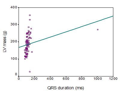
Background: The echocardiographic measurement of left ventricular dimensions and the changes of left ventricular dimensions may be important to assess cardiac as well as the cardiovascular conditions of patients. On the other hand, the QRS duration signifies the time for ventricular depolarization. Aim of the study: The aim of this study was to assess the echocardiographic left ventricular dimensions with several clinical findings among healthy people. Methods: This was an observational cross-sectional study which was conducted in the Department of Cardiology, University Cardiac Centre, Bangabandhu Sheikh Mujib Medical University, Dhaka over a period of 2 years from July 2008 to June 2010. In total 92 apparently healthy people without heart failure (HF) or myocardial infarction (MI) were included as the study population. Among them, 22 were in Referent (QRS duration <100 ms), 40 in Incomplete BBB (QRS duration 100 – 119), and 30 in Complete BBB (≥ 120 ms) groups. Proper written consent was taken from all the participants and this study was approved by the ethical committee of the mentioned university. All data were processed, analyzed, and disseminated by MS Office and SPSS version as per need. Results: In this study, the left ventricular (LV) mass, left ventricular diastolic dimension, septal wall thickness, posterior wall thickness and left atrial size were significantly higher in complete BBB group than those in referent and incomplete BBB group (143.0 ± 30.2 vs. 182.6 ± 37.9 vs. 222.0 ± 61.0, p < 0.001; 4.6 ± 0.3 vs. 4.9 ± 0.3 vs. 5.2±0.4, p< 0.001; 0.9±0.1 vs. 1.0 ± 0.1 vs. 1.1± 0.1, p<0.001; 0.9 ± 0.1 vs. 1.0 ± 0.1 vs. 1.1± 0.1, p<0.001; and 3.3 ± 0.3 vs. 3.6±0.3 vs. 3.7±0.3, p<0.001 respectively). However, fractional shortening and left ventricular ejection fraction were found to decrease significantly with the increase in QRS duration (p < 0.001 and p < 0.001 respectively). In this study the QRS duration was observed to be linearly correlated with LV mass, LV diastolic dimension, septal wall thickness, posterior wall thickness and left atrial size (r=0.577, p<0.001; r=0.480, p<0.001; r=0.583, p<0.001; r=0.521, p< 0.001 and r= 0.418, p<0.001 respectively). However, QRS duration was found to maintain a negative correlation with fractional shortening and LVEF (r =- 0.637, p<0.001 and r =- 0.701, p<0.001 respectively). Conclusion: The present study revealed that longer electrocardiographic QRS duration was correlated with the increase in LV mass, LV diastolic dimensions, septal wall thickness, posterior wall thickness, and left atrial size. The association was most striking in individuals with complete BBB compared with a normal QRS duration. Meanwhile, the presence of prolonged QRS in a patient’s ECG can serve as a bedside clue to the presence of decreased fractional shortening and left ventricular ejection fraction.
Downloads
References
Francia P, Ballac, Paneni F & Volpem M 2007, Left bundle- branch block-pathophysiology, prognosis, and clinical management, Clin. Cardiol, vol.30, pp.110-15.
Murkofsy RL, Danges G, Diamond JA, Mehta D, Schaffer A & Ambrose JA 1998, A prolonged QRS duration on surface electrocardiogram is a specific indicator of left ventricular dysfunction, J Amcoll Cardiol, vol.32, pp. 476-82.
David M, Mirvis & Ary L. Gold berger. Electrocardiography. Brawnwald's Heart disease. A text book of cardiovascular Medicine, Page-149; 8th edition, 2008.
Dhingra R, Ho Nam B, Benjamin EJ, Wang TJ, Larson MG & D' Agostino S 2005, Cross-sectional relations of electrocardiographic QRS duration to left ventricular dimensions: The Framingham Heart study, J Am Coll Cardiol, vol.45, pp. 685-9.
Emkanjoo Z, Esmaeilzadeh M, Hadi NM, Alizadeh A, Tayyebi M & Sadr-ameli MA 2007, Frequency of inter -and intra ventricular dyssynchrony in patient with heart failure according to QRS width, Europace, vol.9, pp.1171-6.
Prinzen FW, Cheriex delhaas T 1995, Asymmetric thickness of the left ventricular wall resulting from asynchronous electric activation: A study in dogs with ventricular pacing and in patients with left bundle branch block, Am Heart J, vol.130, pp.1045-53.
Shenkaman HJ, Pampati V & Khandelwal AK 2002, Congestive heart failure and QRS duration: establishing prognosis study, Chest, vol.122, pp.528-34.
Schneider JF, Thomas HE, Kreger BE, et al. Newly acquired right bundle-branch block: The Framingham Study. Ann Intern Med. 1980; 92:37-44.
Schneider JF, Thomas HE Jr, McNamara PM, Kannel WB. Clinicalelectrocardiographic correlates of newly acquired left bundle branch block: The Framingham Study. Am J Cardiol. 1985; 55:1332-1338.
Schneider JF, Thomas HE, Sorlie P, et al. Comparative features of newly acquired left and right bundle branch block in the general population: The Framingham study. Am J Cardiol. 1981; 47:931-940.
Kreger BE, Anderson KM, Levy D. QRS interval fails to predict coronary disease incidence: The Framingham Study. Arch Intern Med. 1991;151;1365-1368.
Dhingra R, Pencina MJ, Wang TJ, Nam BH, Benjamin EJ & Levy D et al., 2006, Electrocardiographic QRS Duration and the Risk of Congestive Heart Failure, Hypertension, vol.47, pp.861.