THE CHANGING IMMUNOHISTOCHEMICAL PROFILE OF BREAST CARCINOMAS IN NNEWI, SOUTH-EAST NIGERIA: OUR EXPERIENCE
Changing Immunohistochemical Profile of Breast Carcinomas
Abstract
Introduction: GLOBOCAN statistics has shown a rising incidence of breast cancers (BCs) in Nigeria, with previous studies in our region showing predominance of triple negative category. The molecular classification of BCs, as refined in 2015 by the St. Gallen’s consensus, will help prognosticate and better personalize treatment even in resource limited countries.
Objective: The aim of this study was to evaluate immunohistochemically (IHC) status of BCs diagnosed in Nnewi, southeast Nigeria.
Materials and Methods: We analysed all the morphologically diagnosed cases of BCs that were evaluated IHC for hormone receptors (HR) status, HER2 and Ki67 from two histopathology laboratories serving Nnewi and environs over a 6 year period using SPSS software, version 20.
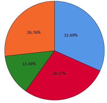
Results: A total of 13275 surgical specimens were received, 2888 of which were solid malignancies and 2485 were breast tissue specimens (974 (39.20%) of which were BCs). BCs accounted for 33.73% of all solid cancers. Only 142 had IHC done on the tissue blocks. The age ranged from 21 to 80 years with modal age in the 5th decade and mean age of 49.90±12.1 years. Majority (59.2%) of the BCs were HR positive and only 17.1% of BCs in women ≤40years were triple negative (TN). HER2 BCs and triple negative breast cancers (TNBCs) were commoner in women aged > 40 years.
Conclusion: There is higher proportion of hormone receptor positive breast cancers in this study compared to previous study in our locality and other parts of the country. The higher proportion of triple negative breast cancers in other studies may be due to pre-analytic factors.
Downloads
References
Falck AK, Fernö M, Bendahl PO, Rydén L. St Gallen molecular subtypes in primary breast cancer and matched lymph node metastases - aspects on distribution and prognosis for patients with luminal A tumours: results from a prospective randomised trial. BMC Cancer 2013, 13:558. DOI: 10.1186/1471-2407-13-558.
Sung H, Ferlay J, Siegel RL, Laversanne M, Soerjomataram I, Jemal A, et al. Global cancer statistics, 2020: GLOBOCAN estimates of Incidence and Mortality worldwide for 36 cancers in 185 countries. CA Cancer J Clin. 2021; 71(3):209-249.
GLOBOCAN 2020: Nigeria-The Global Cancer Observatory march 2021. www.gco.iarc.fr. Accessed 09 May, 2021.
GLOBOCAN 2018: Nigeria-The Global Cancer Observatory march 2021. www.gco.iarc.fr. Accessed 09 May 2021.
Dewan K, Mandal AK. Surrogate molecular classification of breast carcinoma: A classification in need or a dilemma indeed. Oncol J India 2020;4:79-86. DOI: 10.4103/oji.oji_46_19
Vasconcelos I, Hussainzada A, Berger S, Fietze E, Linke J, Siedentopf F, et al. The St. Gallen surrogate classification for breast cancer subtypes successfully predicts tumor presenting features, nodal involvement, recurrence patterns and disease free survival. The Breast. 2016; 29: 181-185. http://dx.doi.org/10.1016/j.breast.2016.07.016.
Esposito A, Criscitiello C, Curigliano G. Highlights from the 14th St Gallen International Breast Cancer Conference 2015 in Vienna: Dealing with classification,prognostication, and prediction refinement to personalize the treatment of patients with early breast cancer. ecancer 2015, 9:518 DOI: 10.3332/ecancer.2015.518.
Lester SC. The Breast. In: Kumar V, Abbas AK, Aster JC (eds). Robbins and Cotrans Pathologic Basis of Diseases. 9th edition. Elsevier Saunders, Philadelphia, USA.2015; 1043-1071.
Ohehe-Yeboah M, Adjei E. Breast cancer in Kumasi, Ghana. Ghana Med J 2012; 46(1):8-13.
Bird PA, Hill AG, Houssami N. Poor hormonal receptor expression in east Africa breast cancer: Evidence of a biologically different disease? Ann Surg Oncol 2008; 15(7):1983-88.
Titiloye NA, Omoniyi-Esan GO, Adisa AO, Komolafe AO, Afolabi OT, Adelusola KA. Breast cancer in Nigeria Cohort: Histopathology, Immunohistochemical profile, and Survival. Postgraduate Medical Journal of Ghana 2013; 2 (2):83-87;
Adani Ifè A, Amégbor K, Doh K, Darré T. Breast cancer in togolese women: immunohistochemistry subtypes. BMC Women’s Health. 2020; 20:261 https://doi.org/10.1186/s12905-020-01130-2;
Ukah CO, Emegoakor C , Anyiam DCD , Onyiaorah IV, Onwukamuche ME, et al. The Immunohistochemical Profile of Breast Cancer in Indigenous Women of Southeast Nigeria. Ann Med Health Sci Res. 2017; 7: 83-87.
Adebamowo CA, Famooto A, Ogundiran TO, Aniagwu T, Nkwodimmah C, Akang EE. Immunohistochemical and molecular subtypes of breast cancer in Nigeria. Breast Cancer Res Treat. 2008; 110:183–188.
Engel KB, Moore HM. Effects of preanalytical variables on the detection of proteins by immunohistochemistry in formalin-fixed, paraffin-embedded tissue. Arch Pathol Lab Med. 2011; 135:537–543;
Pikkarainen M, Martikainen P, Alafuzoff I. The effect of prolonged fixation time on immunohistochemical staining of common neurodegenerative disease markers. J Neuropathol Exp Neurol. 2010; 69:40–52.
Ramos-Vara JA, Miller MA. When tissue antigens and antibodies get along: revisiting the technical aspects of immunohistochemistry—the red, brown, and blue technique. Vet Pathol 2014; 51:42
Grillo F, Bruzzone M, Pigozzi S, Prosapio S, Migliora P, Fiocca R, Mastracci L. Immunohistochemistry on old archival paraffin blocks: is there an expiry date? J Clin Pathol. 2017; 70:988–993
Lundström Y, Lundström P, Popova SN, Lindblom RPF, Alafuzoff I. Detection of changes in immunohistochemical stains caused by postmortem delay and fixation time. Appl Immunohistochem Mol Morphol. 2018. https://doi.org/10.1097/PAI.0000000000 000658.
Report of the Nigeria’s National Population Commission on the 2006 Census. Popul. Dev. Rev. 2007; 33:206-210
WHO Classification of Tumours Editorial Board. Breast tumours. Lyon (France): International Agency for Research on Cancer; 2019. (WHO classification of tumours series, 5th ed.; vol. 2). https://publications.iarc.fr/581.
Elston CW, Ellis LO. Pathological prognostic factors in breast cancer.I. The value of histological grade in breast cancer: experience from a large study with long-term follow-up. Histopathology 1991; 19: 403-410.
Allison KH, Hammond MEH, Dowsett M, McKernin SE, Carey LA, Fitzgibbons PL, et al. Estrogen and Progesterone receptor testing in Breast cancer: ASCO/CAP Guideline Update. J clin Oncol. 2020; 38(12): 1346-1366. DOI: 10.1200/JCO.19.02309.
Wolff AC, Hammond MEH, Allison KH, Harvey BE, Manqu PB, Bartlett JMS, et al. human epidermal growth factor receptor 2 testing in breast cancer: American society of clinical oncology/college of American pathologists clinical practice guideline focussed update. J clin Oncol. 2018; 36(20): 2105-2124. DOI: 10.1200/JCO.2018.77.8738
Trop I, LeBlanc SM., David J, Lalonde L, Tran-Thanh D, Labelle M, et al. Molecular Classification of Infiltrating Breast Cancer: Toward Personalized Therapy. RadioGraphics 2014; 34:1178–1195. DOI: 10.1148/rg.345130049.
Nwafor CC, Keshinro P. Pattern of hormone receptors and human epidermal growth factor receptor 2 status in sub-Saharan breast cancer cases: Private practice experience. Nigerian Journal of Clinical Practice 2015; 18: 553-558.
Gukas ID, Jennings BA, Mandong BM. Clinicopathological features and molecular markers of breast cancer in Jos, Nigeria. WAJM. 2005; 24: 209-213
Chand P, Garg A, Singla V, Rani N. Evaluation of immunohistochemical profile of breast cancer for prognostics and therapeutic use. Niger J Surg. 2018;24: 100-6
Tsang JYS, Tse GM. Molecular Classification of Breast. Adv Anat Pathol. 2020;27:27–35
Kim SW, Roh J, Park CS. Immunohistochemistry for Pathologists: Protocols, Pitfalls, and Tips. Journal of Pathology and Translational Medicine. 2016; 50: 411-418. https://doi.org/10.4132/jptm.2016.08.08
Ramos-Vara JA. Technical Aspects of immunohistochemistry. Vet Pathol. 2005; 42:405–426 ()
Bussolati G, Leonardo E. Technical pitfalls potentially affecting diagnoses in immunohistochemistry. J Clin Pathol 2008; 61:1184–1192. doi:10.1136/jcp.2007.047720
Sengal AT, Haj-Mukhtar NS, Elhaj AM, Bedri S, Kantelhardt EJ, Mohamedani AA. Immunohistochemistry defined subtypes of breast cancer in 678 Sudanese and Eritrean women; hospitals based case series. BMC Cancer. 2017; 17:804 DOI: 10.1186/s12885-017-3805-4
Tiwari S, Malik R, Trichal VK, Nigam RK, Rai A, Balani S, et al. breast cancer: Correlation of molecular classification with clinicopathology. Sch. J. App. Med. Sci., 2015; 3(2G): 1018-1026.
Howlander N, Noone AM, Krapcho M, Miller D, Brest A, Yu M, et al (editors). SEER Cancer statistics Review, 1975-2018. National Cancer Institute. Bethesda, MD. Accessed on october 20, 2021 from https://seer.cancer.gov/csr/1975_2018/,2021.
Shafatujjahan A, Biswas RSR, Ifatujjahan S. Molecular Subtypes of Breast Cancer Patients According to St Gallen Classification. Chattogram Maa-O-Shishu Hospital Medical College Journal. 2020; 19(1):55-58
Galukande M, Wabinga H, Mirembe F, Karamagi C, Asea A. Molecular breast cancer subtypes prevalence in an indigenous Sub Saharan African population. Pan African Medical Journal. 2014; 17:249. doi:10.11604/pamj.2014.17.249.330.
Morris GJ, Naidu S, Topham AK, Guiles F, Xu Y, McCue P, et al. Differences in Breast Carcinoma Characteristics in Newly Diagnosed African-American and Caucasian Patients: A Single-Institution Compilation Compared with The National Cancer Institute’s Surveillance, Epidemiology, and End Results Database. Cancer. 2007;110(4):876–84.
Taminowo MO, Abudu EK, Udo IA, Abdulkareem FB. Histopathological and Immunohistochemical Characteristics of Breast Carcinomas in Uyo, Subtropical Region of Africa. Medical Journal of Zambia. 2019; 46 (2): 100 – 108
Singh LJ, Devi YS, Mohanty S, Nongrum DL. Clinico-Epidemiological and Pathological Characteristics of Breast Cancer in Young Women (<40Years). Ann. Int. Med. Den. Res. 2019; 5(2):MC09-MC14.
Miguel F, Lopes LV, Ferreira E, Ribas E, Pelaez AF, Leal C, et al. Breast cancer in Angola, molecular subtypes: a first glance. ecancer 2017; 11:763 https://doi.org/10.3332/ecancer.2017.763