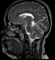
Various presentations of intracranial meningiomas
Abstract
Meningiomas are the most common non-glial tumor of the central nervous system (CNS). There are a number of characteristic imaging features of meningiomas on magnetic resonance imaging (MRI) that allow an accurate diagnosis, however there are a number of atypical features that may be diagnostically challenging. Furthermore, a number of other neoplastic and non-neoplastic conditions may mimic meningiomas.
Downloads
References
Toh CH et al (2008) Differentiation between classic and atypical meningiomas with use of diffusion tensor imaging. AJNR Am J Neuroradiol 29(9):1630–1635
Whittle IR et al (2004) Meningiomas. Lancet 363(9420):1535–1543
O’Leary S et al (2007) Atypical imaging appearances of intracranial meningiomas. Clin Radiol 62(1):10–17.
Buetow MP, Buetow PC, Smirniotopoulos JG. Typical, atypical, and misleading features in meningioma. Radiographics. 1991;11(6):1087–106.
Santelli L, Ramondo G, Della Puppa A, Ermani M, Scienza R, d’Avella D, et al. Diffusion-weighted imaging does not predict histological grading in meningiomas. Acta Neurochir (Wien). 2010;152(8):1315–9 (discussion 9)
Tokumaru A, O’Uchi T, Eguchi T, Kawamoto S, Kokubo T, Suzuki M, et al. Prominent meningeal enhancement adjacent to meningioma on Gd-DTPA-enhanced MR images: histopathologic correlation. Radiology. 1990;175(2):431–3.
Report of brain tumor registry of Japan (1984–2000). Neurol Med Chir (Tokyo). 2009;49 Suppl:PS1-96.
Ko KW, Nam DH, Kong DS, Lee JI, Park K, Kim JH. Relationship between malignant subtypes of meningioma and clinical outcome. J Clin Neurosci Off J Neurosurg Soc Australas. 2007;14(8):747–53.
Wang S, Kim S, Zhang Y, Wang L, Lee EB, Syre P, et al. Determination of grade and subtype of meningiomas by using histogram analysis of diffusion-tensor imaging metrics. Radiology. 2012;262(2):584–92.


