Role of MRI in evaluation of cervical cancer and it’s clinical & histopathological correlation
Abstract
Background: Cervical cancer is one of the leading causes of mortality and morbidity in women. Clinical FIGO staging has been used traditionally but due to its ineffectiveness regarding tumor extent, stromal invasion, distant organ invasion and overstaging of tumors, it is not considered the gold standard for staging of tumor. Use of MRI is now being encouraged for the pre-treatment evaluation of carcinoma cervix. Histopathology remains the most commonly utilized diagnostic tool of cervical cancers. This study was planned with the aim to compare the diagnostic performance of MR Imaging, using histology as the gold standard, with regard to the presence, size and extent of invasive cervical cancers and the detection of metastatic lymph nodes and prognosticates disease outcome and treatment modality and thus reduce the morbidity and mortality associated with the disease. Materials and methods: This study was conducted in the department of Radio diagnosis, Dr. Balasaheb Vikhe Patil Rural Medical College and Dr. Vitthalrao Vikhe Patil Pravara Rural Hospital, Loni BK, 413736 during the period of June 2021 to June 2022. It was a retrospective study, 30 cases of clinically suspected or diagnosed as carcinoma cervix by biopsy and Pap smear, referred to the department of Radio diagnosis. Imaging was done with 3 Tesla Philips Ignesia.
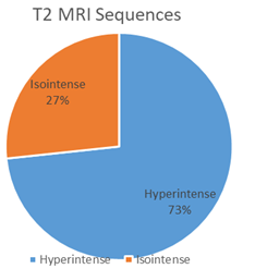
Results: Among the study population, the mean age observed was 55.62 ± 15.0 years, with BMI 20.65 ± 3.29 kg/m2 and majority belonged to lower socio-economic status. The common risk factors associated with Carcinoma Cervix was multiparity (74%) and the most common presenting complaint was foul smelling vaginal discharge observed in 58% cases. 5 cases out of 30 that were diagnosed as IA on clinical examination and on MRi they were staged as IB, IIA and IIB on MRI. Rest of the cases were staged as shown in the table and the results were Statistically significant. (p value<0.007304)
Conclusion: Carcinoma cervix is primarily staged clinically as per FIGO guidelines but MRI can modify treatment options and may provide clinically important prognostic information not available from current FIGO staging. MRI also has the potential to be used as diagnostic tool for cervical cancer as it correlates strongly with histopathology.
Downloads
References
- Zhang S, Xu H, Zhang L, Qiao Y. Cervical cancer: Epidemiology, risk factors and screening. Chin J Cancer Res 2020;32(6):720-728. doi: 10.21147/j.issn. 1000-9604.2020.06.05.
- Mishra GA, Pimple SA, Shastri SS. An overview of prevention and early detection of cervical cancers. Indian J Med Paediatr Oncol 2011; 32:125-32.
- Yogaraj, Kumaran. Association between MRI Findings and Histopathological Examination in Carcinoma Cervix: A Retrospective Study. International Journal of Anatomy, Radiology and Surgery. 2021 Apr, Vol-10(2): RO61-RO64.
- Shweel MA, Abdel-Gawad EA, Abdelghany HS, Abdel-Rahman AM, Ibrahim EM. Uterine cervical malignancy: diagnostic accuracy of MRI with histopathologic correlation. J Clin Imaging Sci. 2012; 2:42. PMID: 22919556.
- Morimura Y, Soeda S, Hashimoto T, Takano Y, Ohwada M, Yamada H, et al. The value of pre-operative diagnostic procedures for cervical involvement in uterine corpus carcinoma. Fukushima J Med Sci. 2000;46(1-2):1-11.
- D. Amitha Kumari, Ananthalakshmi Paga. Comparative study of MRI staging vs figo staging of carcinoma cervix. International Journal of Contemporary Medical Research 2017;4(5):1196- 1198.
- Giuliano Rigon, Cristina Vallone, Andrea Starita, Marco Flavio Michele Vismara, Pasquale Ialongo, Lorenza Putignani, Fabrizio Signore. Open Journal of Radiology. 2012; 2:14-21.