Clinical outcome, risk factors and angiographic evaluation of inferior myocardial infarction with or without right ventricular involvement
Abstract
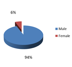
Introduction: Assessment and quantification of right ventricular function is difficult and challenging. Nevertheless, an understanding of right ventricular function may be useful in the management of patients with an inferior acute MI that involves the right ventricle. The extent of involvement of the right ventricle varies in different series and angiographic analysis of the right ventricle can shed light on the severity of the disease. Objective: To assess the clinical outcome, risk factors and angiographic evaluation of inferior myocardial infarction with or without right ventricular involvement. Methods: This was a prospective observational comparative study carried out in National Institute of Cardiovascular diseases (NICVD) Dhaka, during the period from October 2010 to June 2011. One hundred (100) patients both group included. Among the patients we selected 50 patients with RV myocardial involvement (Group I= ST elevation >1mm in V4R) and 50 patients without RV myocardial involvement (Group II= ST isoelectric or depression in V4R). Patients admitted in CCU with AMI (inferior) fulfilling the inclusion and exclusion criteria were included in the study. According to ECG finding in right precordial lead V4R, patients were categorized into two groups. Echocardiography was done in all the patients within 24 hours of onset of chest pain. After 7 to 10 days all patients had undergone coronary angiography and then evaluation done. All data were recorded in preformed data sheet and analysis was done by computer based on SPSS program. P-value less than (< 0.05) considered as statistically significant. Results: One hundreds (100) patients were both group included. The mean age of Group I and Group II patients were (54.5±11.2 vs 54.5±11.2 years. P=0.08). The highest number of patients was in the age group (50-59) years. Majority of patients were male 94% vs. 92% respectively. On admission, chest pain was the most common presenting compliant both Groups (100% vs. 98%), breathlessness (60% vs. 24%), nausea (54% vs. 58%), vomiting (90% vs. 86%), sweating (90% vs. 98%), syncope (60% vs. 20%) and dizziness (70 vs. 28%) between Group I and Group II respectively. CAG report of coronary artery showed that 68% of the patients had lesion in proximal of the RCA in Group I. In middle of the RCA had 6% vs. 44% of patients in between Group I and Group II. The difference was statistically significant (p<0.001). There were no patients in distal part of RCA in Group I and had 36% of patients in Group II. LCX had 34% vs.26% in Group I and Group II respectively and (p=0.38). Among the studied patients, the most important frequent complications were hypotension followed by sinus bradycardia, cardiogenic shock, arrhythmias, acute LVF and cardiac arrest and death.
Downloads
References
Ali,M .2006 , “Coronary heart diseases : Need for epidemiological studies and guideline for south Asians,” Bangladesh Heart J. vol.21(2) ,p.1.
Fuster ,V 1999, “ Epidemic of cardiovascular disease and stroke : Three main challenges ,” Circulation ,vol. 99,pp.1132-1137.
Reddy, K.S. Yusuf, S 1998, ‘Emerging epidemic of cardiovascular disease in developing countries’, Circulation, Vol.97, pp. 596-601.
Yusuf, S. Reddy, S. Ounpuu, s & Anand, s 2001, ‘Global burden of diseases, part I: general considerations, the epidemiologic transition, risk factors and impact of urbanization’, Circulation, vol.104, pp. 2746-2753.
Yusuf, S. Reddy, S. Ounpuu, s & Anand, s 2001, ‘Global burden of diseases, part II: variations in cardiovascular diseases by specific ethnic groups and geographic and preventive strategies’, Circulation, vol.104, pp. 2855-2864.
Chockalingam, A. Gnanavelu, G. Subramaniam, T . Dorairajan, S and Chockalingam V, 2005 ,’ Right ventricular myocardial infarction .’ Angiology, vol . 56, pp. 371-376.
Majumder , A.A.S , Haque ,A ,Haque , M , Haque ,SA &Chowdhury , S 1996, Right ventricular involvement in acute myocardial infarction : Course ,complications , and management –an experience at a teaching Hospital’, Journal of institute of Postgraduate Medicine & Research ,vol. 11 ,pp. 22-25 .
Kurtulus, O. Bulent, B. & Altunkeser ,B 2005 , ‘New approach in identification of Rt ventricular myocardial infarction and proximal Rt coronary artery lesion ,’ Chest , vol . 126, pp .319 -325.
Bergsma, E and Mircade, N 2003, “ECG evalution in all acute myocardial infarction ,” Lancet , vol . 361, pp. 847- 858.
Burgess, M.I. Bright, RJ. Ray, SG. 2002, ‘Echocardiographic evaluation of Right ventricular function,’ Euro J Echocardiography ‘vol. 3, pp. 252-262.
Shawkat , AH and Assad , M 2000,’ Right ventricular infarction Diagnosis and Treatment ‘,Clinical Cardiology ,vol . 23 ,pp . 473 -482.
Falk, E. Shah, P.K. Fuster, V 2008, “Atherothrombosis : Role of inflammation,” ‘In Fuster ,V . Walsh, R. A. O’Rourke, R. A. Wilson, P. P. (eds) ,Hurst’s The Heart , 12th edn , McGraw – Hill , New York ,USA,PP.1235 -1240 .
Jamaluddin ,M . Haque , KMHSS . Chowdhury , AHK. & Zaman ,MA .1997 , ‘ Short term(1 month) Prognosis of patients of Acute Myocardial Infarction Receiving Thrombolytic Therapy ,’Bangladesh Heart Journal ,vol. 12 (2) , pp. 54-58 .
Baigrie, R.S. Haq, A . Christopher, D. Morgan, Rakowski, H. Drobac, M. Mclaughlin , P. 1983 . “The Spectrum of Right Ventricular Involvement in Inferior Wall Myocardial Infarction: A Clinical, Hemodynamic and Noninvasive Study”, Jam,coll. cardio, Vol .1(6), Pp.1396-404
Assali, A.R. Herz, M.V. Adler, B. & Samul ,S 1999, “Electrocardiographic criteria for predicting the culprit artery in inferior wall acute myocardial infarction”, American journal of cardiology , vol ,81 , pp.87-89.
Braat, S.H. Karel, H. Pedro, Brugada, p. & Weallens ,J.J. 1984 ,’ Value of lead V4R for Recognition of the infarct coronary artery in Acute inferior wall myocardial infarction’ , American journal of cardiology, vol.53 ,pp.1538-1541.
Hurst, J.W. 1998, “Comments about the Electrocardiographic signs of right ventricular infarction” Clin Cardiol, vol .21(4) pp .289-91.
Islam, M.N. Ali, M.A. Ali, M 2004, “Spectrum of cardiovascular disease: the current scenario in Bangladesh,” Bangladesh Heart J, vol. 19, pp.1-7.
Khan, S. Kundi, A. Sharief 2004, “Prevalence of right ventricular myocardial infarction in patients with acute inferior wall myocardial infarction.” Int J Clin Pract ; 58(4): 354-7.
Goldstein, J.A. 2002, “Pathophysiology and management of right heart ischemia.” J Am Coll Cardiol ,vol.40 , pp .841–53.
Khalil, M. Hossain, M. Ahmed, C.M. Jalaluddin, M. Majumder, A.A.S 1997, “Precordial ST segment depression in acute inferior myocardial infarction: A marker of concomitant anterior ischemia,” Bangladesh Heart J, vol. 12 ,pp. 8-10 .
Arcy, B.D. Nanda, N.C 1982, “Two-dimensional echocardiographic features of right ventricular infarction”, Circulation, vol .65 pp. 167-173.
Mehta , S .R. Eikelboom ,J .W . Natarajan , M .K. Diaz ,R. Yi C, Gibbons ,R.J .Yusuf ,S. 2001 , “ Impact of right ventricular involvement on mortality and morbidity in patients with inferior myocardial infarction ,” J Am Coll Cardiol. Vol. 37, pp .37– 43.