The validity of plain lumber vertebral X-Rays in diagnosing osteoporosis in elderly-An age-based approach
Abstract
Background: The diagnosis of osteoporosis relies on the quantitative assessment of BMD, which is currently considered the best predictor of osteoporotic fractures. Early diagnosis is the key for appropriate osteoporosis management. Although common, osteoporosis can be clinically silent, and without prevention and screening, the costs of osteoporotic fracture–related morbidity and mortality will burden healthcare systems, especially in developing countries.
Objective: To assessed the validity of plain radiography in diagnosing osteoporosis in elderly women.
Methods: A retrospective, cross-sectional, observational hospital-based study conducted at the Department of Ortho-Surgery, Patuakhali Medical College Hospital, Patuakhali, Bangladesh from June 2019 to July 2022. One hundred Seventy (170) female patients between the ages of 40 to 83 years were referred to the orthopedic department in PKMCH. These women were found to have features of osteopenia in lumber vertebrae plain radiography. The participants then categorized into two groups. Group A (n=101) are those who are younger than 65 years and group B (n=69) are those who are 65 years and older. The two groups underwent a quantitative ultrasound bone densitometry. Correlations between plain radiography parameters and QUS were calculated. Osteoporosis was diagnosed by QUS T-score ≤ –2.5 at the lumber vertebra.
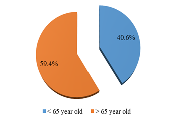
Results: Total 170 patients were included. The mean age of the participants was 63.5±6 years old with the minimum age was 40 years and the maximum age was 83 years. The most common population aged more than 63 years old, group A who are less than 65 years of age were 101 participants (59.4%), while those 65 years and old were 69 (40.6%). The participants in both groups have showed features of osteopenia in their plain lumbar vertebral X-rays. By QUS; in group A: 2 patients (1.9%) were found to have a normal bone mineral density (T score = >-1 SD), 47 patients (46.5%) were osteopenic (T score between -1 and -2.5 SD), while 52 patients (51.4%) were osteoporotic (T score = <-2.5 SD), in group B: 3 patients (4.3%) were found to have a normal bone mineral density (T score =>-1 SD), 3 patients (4.3%) were osteopenic (T score between -1 and -2.5 SD), while 63 patients (91.3%) were osteoporotic (T score =<-2.5 SD). Also when we performed Fisher’s Exact test we found a significant difference in the validity of X rays as compared to QUS bone densitometry between the two groups, in Group A. The difference between X-ray and quantitative ultrasound bone densitometry was significant (p = 0.000000006 at p > 0.05), and was not significant in Group B (p = 0.491 at p >0.05).
Conclusion: Plain radiography can provide reliable method for diagnosis of osteoporosis in women with a higher risk for fragility fractures (≥65 years) especially in primary healthcare and sittings with limited resources.
Downloads
References
Eastell R. Treatment of postmenopausal osteoporosis. N Engl J Med. 1998; 338:736–746.
Seeman, E. (2003). Osteoporos Int 14(Suppl 3), 2. https://doi.org/10.1007/s00198-002- 1340-9.
Assessment of fracture risk and its application to screening for postmenopausal osteoporosis. World Health Organ Tech Rep Ser. 1994; 843:1–129.
Grigoryan, M., Guermazi, A., Roemer, F. W., Delmas, P. D., & Genant, H. K. (2005). Recognizing and reporting osteoporotic vertebral fractures. In The Aging Spine (pp. 22- 30). Springer, Berlin, Heidelberg.
Fink, H. A., Milavetz, D. L., Palermo, L., Nevitt, M. C., Cauley, J. A., Genant, H. K., & Ensrud, K. E. (2005). What proportion of incident radiographic vertebral deformities is clinically diagnosed and vice versa? Journal of bone and mineral research, 20(7), 1216- 1222.
Panda, A., Das, C.J., & Baruah, U. (2014). Imaging of vertebral fractures. Indian journal of endocrinology and metabolism, 18(3), 295.
Glüer CC, Wu CY, Jergas M, Goldstein SA, Genant HK. Three quantitative ultrasound parameters reflect bone structure. Calcif Tissue Int. 1994; 55:46–52.
Hayes WC, Piazza SJ, Zysset PK. Biomechanics of fracture risk prediction of the hip and spine by quantitative computed tomography. Radiol Clin North Am. 1991; 29:1–18.
Kaufman JJ, Einhorn TA. Ultrasound assessment of bone. J Bone Miner Res. 1993; 8:517–525.
Njeh CF, Boivin CM, Langton CM. The role of ultrasound in the assessment of osteoporosis: a review. Osteoporos Int. 1997; 7:7–22.
McCullagh, C. D., McCoy, K., Crawford, V. L. S., & Taggart, H. (2003). How reliable is a radiological report on osteoporosis in diagnosing low bone density? The Ulster medical journal, 72(1), 34.
Scane, A. C., Masud, T., Johnson, F. J., & Francis, R. M. (1994). The reliability of diagnosing osteoporosis from spinal radiographs. Age and Aging, 23(4), 283-286.
Masud, T., Mootoosamy, I., McCloskey, E. V., O'Sullivan, M. P., Whitby, E. P., King, D., & Spector, T. D. (1996). Assessment of osteopenia from spine radiographs using two different methods: the Chingford Study. The British journal of radiology, 69(821), 451-456.
Garton, M. J., Robertson, E. M., Gilbert, F. J., Gomersall, L., & Reid, D. M. (1994). Can radiologists detect osteopenia on plain radiographs?. Clinical radiology, 49(2), 118- 122.
Harris, W. H., & Heaney, R. P. (1969). Skeletal renewal and metabolic bone disease. New England Journal of Medicine, 280(4), 193-202.
Guglielmi, G., Balzano, R. F., & Cheng, X. (2018). What is changed in the diagnosis of osteoporosis: the role of radiologists. Quantitative imaging in medicine and surgery, 8(1), 1.
Mora,S., & Gilsanz ,V. (2003). Establishment of peak bone mass. Endocrinology and Metabolism Clinics, 32 (1), 39-63.
Resnick, N. M., & Greenspan, S. L. (1989). Senile'osteoporosis reconsidered. Jama, 261(7), 1025-1029.
Epstein, D. M., Dalinka, M. K., Kaplan, F. S., Aronchick, J. M., Marinelli, D. L., & Kundel, H. L. (1986). Observer variation in the detection of osteopenia. Skeletal radiology, 15(5), 347-349.
Espeland, A., Korsbrekke, K., Albrektsen, G., & Larsen, J. L. (1998). Observer variation in plain radiography of the lumbosacral spine. The British journal of radiology, 71(844), 366-375.