Clinical profile of myopia in adult patients at a rural tertiary care hospital.
Abstract
Introduction: Myopia, also known as short sight is that diopteric condition of eye in which, with accommodation at rest, incident parallel rays come to a focus anterior to the retina.[1] The person can see near objects more clearly than distant ones and are called “short-sighted”. The prevalence of simple myopia and high myopia (degenerative myopia) are increasing globally at an alarming rate, with significant increases in the risks for vision impairment from pathologic conditions associated with high myopia, including retinal damage, cataract and glaucoma. We conducted the study to find out the percentage of mild / moderate/severe myopia in adult patients at a rural tertiary care hospital.
Aims and Objectives:To study the clinical profile of Myopia in patients more than 20 years at a rural tertiary care hospital.
Materials and Methods: An Observational, Descriptive Cross-Sectional Hospital based study was conducted at a tertiary care hospital. A total of 100 patients with Myopia were screened and evaluated. We studied the myopic patients above 20 years attending to our hospital OPD. Family history and information about the risk factors was obtained by using pre structured proforma. Snellen’s chart was used to record the unaided visual acuity and best corrected visual acuity. Color vision was assessed by Ischihara pseudo isochromatic color plates. With the help of retinoscopy, refractive errors were determined. Spherical equivalent of refraction (SER) was calculated . Central corneal thickness was calculated by Pac Scan Pachymeter. Axial length was calculated by A Scan biometry. Dilated fundus examination was done by Direct and indirect ophthalmoscope. Results were analyzed using suitable statistical tests. Grading of myopia was done as Mild Myopia(<3D), Moderate Myopia (3-6 D), Severe Myopia (>6 D) and Pathological myopia with fundus changes.
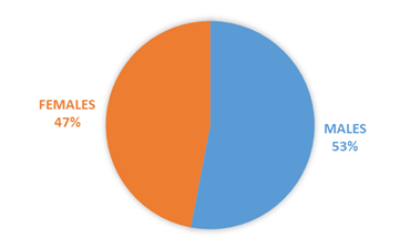
Results: The mean age of the study population was 32.98 ± 15.77 years with 53% (53) males and 47% (47) females. The mean SE was -2.66 ± 3.02 D. 66% (66) patients had mild myopia, 23% (23) patients had moderate myopia,7% (7) patients had severe myopia while 4% (4) patients had pathological myopia with fundus changes.
Conclusion: Occurrence of mild myopia is most common followed by moderate, severe, and pathological myopia which is the least common type.
Downloads
References
Sihota, R.; Tandon, R. Parson’s disease of the eye. 23rd ed. New Delhi: Elsevier; 2003.
Cooper, J; Tkatchenko, A A Review of Current Concepts of the Etiology and Treatment of Myopia
Vision Impairment and blindness World Health Organization. World Health Organization; Available from: https://www.who.int/news-room/fact-sheets/detail/blindness-and-visual-impairment.
Gwiazda J. Treatment options for myopia. Optometry and vision science: official publication of the American Academy of Optometry. 2009 Jun;86(6):624.
Khurana AK. Theory and Practice of Optics and Refraction 4th ed India: RELX India Private Limited.
Dutta LC, Dutta NK. Modern ophthalmology. New Delhi, India: Jaypee Bros.; 2005.
Mehta R, Bedi N, Punjabi S. Prevalence of myopia in medical students. Indian J Clin Exp Ophthalmol. 2019 Jul;5(3):322-5.
Fan Q, Wang H, Jiang Z. Axial length and its relationship to refractive error in Chinese university students. Contact Lens and Anterior Eye. 2022 Apr 1;45(2):101470.
Divya K, Ganesh MR, Sundar D. Relationship between myopia and central corneal thickness–A hospital based study from South India. Kerala Journal of Ophthalmology. 2020 Jan 1;32(1):45.
Solu T, Baravaliya P, Patel I, Savaliya C, Golakiya B. Correlation of central corneal thickness and axial length in myopes, emmetropes, and hypermetropes. International Journal of Scientific Study. 2016;3(12):206-9.
Fam HB, How AC, Baskaran M, Lim KL, Chan YH, Aung T. Central corneal thickness and its relationship to myopia in Chinese adults. British journal of ophthalmology. 2006 Dec 1;90(12):1451-3.


